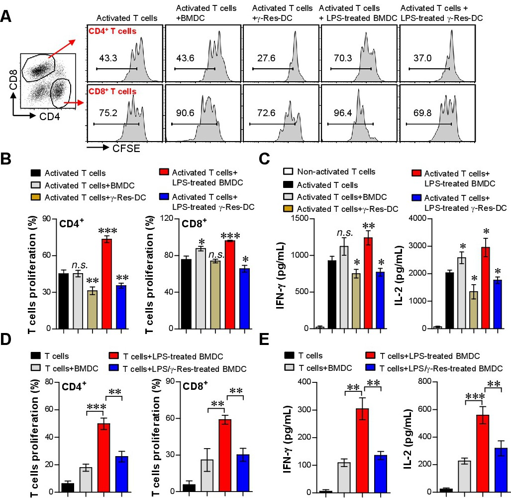
Fig. 9. Inhibitory effect of γ-Res-treated BMDCs and DCs generated by γ-Res on proliferation and activation of T cells. A-C) T cells isolated from the splenocytes of BALB/c and stained with CFSE. The stained T cells were cultured in a 48-well plate coated with anti-CD3 (0.5 μg/mL) and anti-CD28 Abs (0.5 μg/mL). BMDCs and γ-Res-DCs were stimulated with or without LPS for 1 h and co-cultured with the T cells. The ratio between the DCs and T cells was 0.5:1. After 3 days of co-culturing, the cells were harvested and stained with anti-CD4 and anti-CD8 Abs. B) The proliferation in activated-CD4+ and CD8+ T cells was analyzed using flow cytometry. D) Culture mediums were analyzed for the production of IFN-γ and IL-2 cytokines using ELISA. D, E) BMDCs were stimulated with LPS in the absence or presence of γ-Res (30 μg/mL) for 24 h and then co-cultured with T cells isolated from BALB/c for 3 days. The proliferation of T cells and cytokines were measured as described above. All results are representative of three independent experiments. All bar graphs show the means ± SD of three samples per group. *p<0.05, **p<0.01, or ***p<0.001.